
Conjunctival Nevus Surgery in Izmir, Turkey
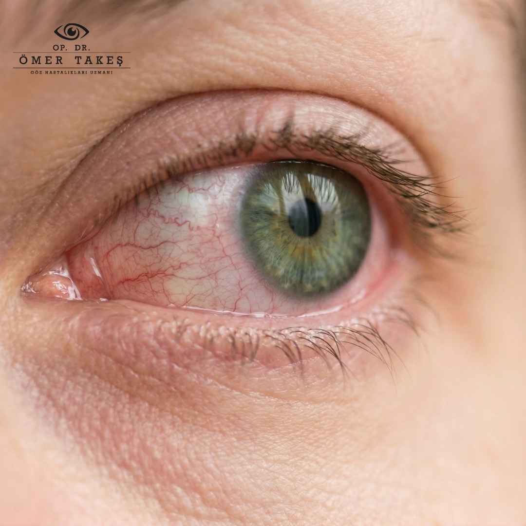
A conjunctival nevus is a pigmented lesion that forms on the conjunctiva, the thin and transparent layer covering the white part of the eye and the cornea. It typically appears in shades of brown, yellow, or pink. While it can be present from birth, it may also develop during adolescence or early adulthood. Most nevi are benign and do not require treatment unless they grow or change in color. However, because there is a rare risk of malignant transformation, regular monitoring is important.
Symptoms and Clinical Features of Conjunctival Nevus
A conjunctival nevus usually does not cause significant symptoms. However, in some cases, the following may be observed:
-
Visible and sometimes cosmetically disturbing spot on the eye surface
-
Flat, brown or black lesions on the white part of the eye
-
Usually painless and do not affect vision; mild irritation may occur rarely
-
Irregular borders, darker pigmentation, or wide distribution may raise concern for malignancy
-
Primary Acquired Melanosis (PAM) typically occurs in middle-aged individuals and may be associated with skin melanomas
Types of Conjunctival Nevus
-
Primary Conjunctival Melanosis
Congenital or genetic, often seen in multiple locations and symmetric. Low risk of malignancy. -
Primary Acquired Conjunctival Melanosis (PAM)
Appears in adulthood with irregular, dark pigmentation. Carries a risk of developing into malignant melanoma, especially if atypical cells are present. -
Congenital Melanosis Oculi
Present at birth, usually unilateral, and appears similar to a nevus. -
Nevus
Benign structure with low malignancy potential. Usually stable, but any change in size or color should be monitored. -
Secondary Conjunctival Melanosis
May occur due to chronic inflammation, trauma, medication use, or systemic diseases.
How Is Conjunctival Nevus Diagnosed?
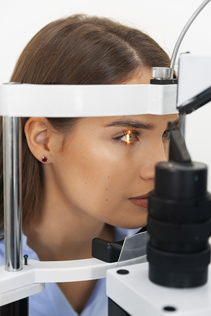
Conjunctival nevus is usually diagnosed during a routine eye examination. An experienced ophthalmologist assesses the size, shape, color, and location of the lesion. If necessary, advanced imaging techniques like ultrasound biomicroscopy or optical coherence tomography (OCT) may be used to evaluate malignancy risk. During clinical examination, the extent and appearance of pigmentation are assessed. The depth and spread of the lesion are evaluated via slit-lamp biomicroscopy. A biopsy is performed if the lesion appears atypical or malignancy is suspected. Histopathological analysis is important for identifying cellular atypia and the risk of malignant melanoma.
What Are the Treatment Options for Conjunctival Nevus?
Most conjunctival nevi do not require treatment. However, in certain cases, treatment may be necessary based on the lesion type, size, and malignancy risk:
-
Monitoring
Regular follow-up may be sufficient for benign nevi or primary melanosis, provided no changes occur. -
Cosmetic Reasons
The lesion may be removed upon patient request for cosmetic concerns. -
Suspicion of Malignancy
If the nevus shows rapid growth, irregular borders, or changes in color, a biopsy, surgical excision, cryotherapy, or radiotherapy may be necessary. Suspicious or fast-growing lesions should be excised and pathologically examined. Surface reconstruction may be required after excision. In some cases, laser or cryotherapy may be used for pigmented lesions. -
Symptomatic Cases
Rarely, a nevus may cause irritation or other discomfort requiring treatment.
Surgical removal is usually performed under local anesthesia and is considered a safe procedure. After excision, the nevus is sent for pathological analysis.
Why Is Regular Monitoring of Conjunctival Nevus Important?
Although the risk of a conjunctival nevus developing into malignant melanoma is very low, it cannot be entirely ruled out. Therefore, pigmented eye lesions should be evaluated by an ophthalmologist to exclude malignancy. Most eye nevi are benign, but those with progression risk should be monitored. An annual eye examination is recommended to follow any changes in the nevus.
Trusted Treatment with Op. Dr. Ömer TAKEŞ
At our clinic located in Alsancak, Izmir, we use state-of-the-art technology for the diagnosis and treatment of conjunctival nevus and other eye conditions. Op. Dr. Ömer TAKEŞ provides personalized and professional care with extensive expertise in his field.
If you notice any spot or abnormal appearance in your eye, we invite you to visit our clinic for a comprehensive evaluation.